Close Menu
  • News
  • Medical
  • Technology
  • Nanomaterials
  • Research
  • Blog
    • Nasiol.com
  • Contact
    • Tech7685@gmail.com
What's Hot

Rapid nanoparticle simulations could boost efforts to combat air pollution

June 1, 2025

New tool reveals how DNA nanostructures interact with cell membranes

June 1, 2025

New insights show universal applicability of carbyne as a sensor

May 31, 2025
Facebook X (Twitter) Instagram
Nanotech – Nanomaterials | Medical | Research | News Stories Updated Daily Nanotech – Nanomaterials | Medical | Research | News Stories Updated Daily
  • News
  • Medical
  • Technology
  • Nanomaterials
  • Research
  • Blog
    • Nasiol.com
  • Contact
    • Tech7685@gmail.com
Facebook X (Twitter) Instagram
Nanotech – Nanomaterials | Medical | Research | News Stories Updated Daily Nanotech – Nanomaterials | Medical | Research | News Stories Updated Daily
Home»News»Upgraded atomic force microscope captures 3D images of calcite dissolving
News

Upgraded atomic force microscope captures 3D images of calcite dissolving

September 7, 2024No Comments4 Mins Read
Facebook Twitter Pinterest Telegram LinkedIn Tumblr WhatsApp Email
Upgraded atomic force microscope captures 3D images of calcite dissolving
Share
Facebook Twitter LinkedIn Pinterest Telegram Email
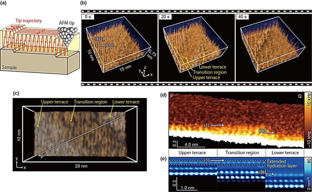
High-speed 3D-SFM imaging (a) Imaging principle of 3D-SFM. (b) 3D-SFM images obtained near the calcite step edge during its dissolution at 5 s / 3D image. (c) Another example of a 3D-SFM image obtained at 1.6 s/3D image. (d) Vertical cross-sectional image obtained over the terrace and transition region. Credit: Nano Letters (2024). DOI: 10.1021/acs.nanolett.4c02368

Understanding the dissolution processes of minerals can provide key insights into geochemical processes. Attempts to explain some of the observations during the dissolution of calcite (CaCO3) have led to the hypothesis that a hydration layer forms, although this has been contested.

Hydration layers are also important as they play a role in a number of processes including adhesion, corrosion and wetting, as well as the folding, stability and recognition of proteins.

Now researchers led by Kazuki Miyata, Adam S. Foster and Takeshi Fukuma at the Nano Life Science Institute (WPI-NanoLSI) at Kanazawa University in Japan have successfully upgraded their atomic force microscope to retrieve imaging data with the time and spatial resolution needed to obtain 3D structure images that provide direct evidence of a hydration layer forming during the dissolution of calcite.

The research is published in the journal Nano Letters.

The hypothesis of a hydration layer forming during the dissolution of calcite was prompted by simulations of the process, which pointed to the production of a Ca(OH)2 layer across “transition regions” as calcite dissolves.

Despite being unstable in the bulk or on flat terraces, Ca(OH)2 can appropriate some stability from step edge structures, although the mechanism behind this is not well understood.

This could explain the stability of the Ca(OH)2 next to the step edges but since the transition regions observed in experiments span several nanometers, the authors had posited the possibility that the Ca(OH)2 acquires its stability through indirect interactions with the step by means of a hydration structure.

However, as the researchers point out in their report, hydration effects remain “poorly understood” as techniques for imaging changes in solid–liquid interfacial structures are lacking.

Atomic force microscopy upgrade captures 3D images of calcite dissolving
A model of the formation and stabilization mechanism of the transition region. Credit: Nano Letters (2024). DOI: 10.1021/acs.nanolett.4c02368

Atomic force microscopy (AFM) obtains high resolution images by using a nanoscale cantilever to feel the surface a little like the needle of a record player feels the grooves in vinyl. However, despite a huge step change in the rate of image acquisition that can be achieved with the invention of high-speed (HS) AFM, AFM has still suffered a little from a trade-off between speed and spatial resolution.

See also  Molybdenene—the 'metallic' relative of graphene

Efforts to apply it to the study of dissolution processes are also hampered because the tool is designed to scan the topologies and interactions across 2D surfaces, and dissolution of minerals involves 3D structural changes.

Previous work had expedited the higher resolution “frequency modulated” (FM) AFM so that the image acquisition time was reduced from a minute to just 0.5 s/frame. This upgrade allowed the authors to image the transition region from which they inferred the presence of a hydration layer, but some extrapolation was required to extract 3D structure information from comparison of the 2D-AFM data to 3D simulation, leaving some to doubt the conclusions.

Modifications of AFM to extract 3D force data using AFM have previously been demonstrated, although once again, despite some improvements to speed things up, to about 1 minute/frame the image acquisition time had remained prohibitive for observing dynamic processes.

The authors get around all these drawbacks by combining the HS-FM-AFM with 3D-SFM. This involved increasing the bandwidth of their 3D-SFM while maintaining a force resolution of 10-100nN, fast synchronization of the signals in the lateral scanning and third dimension, and fast recording of the cantilever frequency shifts. With these in place, the researchers were able to capture 3D-SFM images in just 1.6 s/frame. They used the approach to image the dissolution of calcite.

“The HS-3D-SFM images produced in the present work clearly show the 3D distribution predicted by the simulations, thus supporting the existence of an extended hydration layer,” they point out in their report.

Provided by
Kanazawa University


See also  Fabricating 2D Materials Through Chemical Vapor Deposition


Source link

atomic calcite Captures dissolving Force images microscope Upgraded
Share. Facebook Twitter Pinterest LinkedIn Tumblr Email

Related Posts

Rapid nanoparticle simulations could boost efforts to combat air pollution

June 1, 2025

New tool reveals how DNA nanostructures interact with cell membranes

June 1, 2025

New insights show universal applicability of carbyne as a sensor

May 31, 2025

A new molecular model of bilayer graphene with higher semiconducting properties

May 31, 2025

5 Nanomaterial Innovations That Didn’t Deliver (Yet)

May 30, 2025

Scientists identify new 2D copper boride material with unique atomic structure

May 30, 2025

Comments are closed.

Top Articles
News

High-Speed AFM Advancements for Protein Dynamics Research

News

Nanomedicine research aims to transform treatment of aortic aneurysms

News

Market Trends and Growth Potential

Editors Picks

Rapid nanoparticle simulations could boost efforts to combat air pollution

June 1, 2025

New tool reveals how DNA nanostructures interact with cell membranes

June 1, 2025

New insights show universal applicability of carbyne as a sensor

May 31, 2025

A new molecular model of bilayer graphene with higher semiconducting properties

May 31, 2025
About Us
About Us

Your go-to source for the latest nanotechnology breakthroughs. Explore innovations, applications, and implications shaping the future at the molecular level. Stay informed, embrace the nano-revolution.

We're accepting new partnerships right now.

Facebook X (Twitter) Instagram Pinterest
Our Picks

MIT Physicists Transform Pencil Lead Into Electronic “Gold”

November 6, 2023

Lined-up quantum dots become highly conductive

August 12, 2023

Breakthrough in Weather Monitoring with New Low-Energy Anemometer

July 2, 2024

Subscribe to Updates

Get the latest creative Nano Tech news from Elnano.com

© 2025 Elnano.com - All rights reserved.
  • Contact
  • Privacy Policy
  • Terms & Conditions

Type above and press Enter to search. Press Esc to cancel.

Cleantalk Pixel