Close Menu
  • News
  • Medical
  • Technology
  • Nanomaterials
  • Research
  • Blog
    • Nasiol.com
  • Contact
    • Tech7685@gmail.com
What's Hot

Dual-stage monitoring technique for nanocomposites can streamline manufacturing and property tracking

May 11, 2025

Probing the molecular mechanisms of metastasis

May 10, 2025

AI-powered electronic nose detects diverse scents for health care and environmental applications

May 10, 2025
Facebook X (Twitter) Instagram
Nanotech – Nanomaterials | Medical | Research | News Stories Updated Daily Nanotech – Nanomaterials | Medical | Research | News Stories Updated Daily
  • News
  • Medical
  • Technology
  • Nanomaterials
  • Research
  • Blog
    • Nasiol.com
  • Contact
    • Tech7685@gmail.com
Facebook X (Twitter) Instagram
Nanotech – Nanomaterials | Medical | Research | News Stories Updated Daily Nanotech – Nanomaterials | Medical | Research | News Stories Updated Daily
Home»News»New microscope shows live imaging of nanoscale biological process for the first time
News

New microscope shows live imaging of nanoscale biological process for the first time

November 19, 2024No Comments4 Mins Read
Facebook Twitter Pinterest Telegram LinkedIn Tumblr WhatsApp Email
New microscope shows live imaging of nanoscale biological process for the first time
Share
Facebook Twitter LinkedIn Pinterest Telegram Email
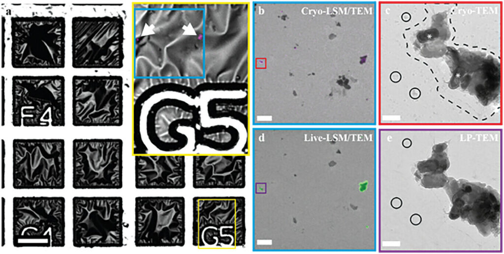
Correlative cryo-/liquid phase imaging of graphene liquid cells. Credit: Advanced Functional Materials (2024). DOI: 10.1002/adfm.202416938

In Nijmegen, Netherlands, researchers have installed the world’s first microscope capable of live imaging of biological processes in such detail that moving protein complexes are visible. This new microscopic technique was developed by researchers led by Nico Sommerdijk from Radboud university medical center. As a demonstration of this innovative technique, Sommerdijk is now showcasing how arterial calcification begins.

Previously, researchers faced a choice. They could use a microscope to examine material in detail down to the molecular level, but only in frozen, motionless samples. Alternatively, they could observe living, moving material, but with far less detail. Radboudumc researchers have developed a technique that combines both approaches. This opens up various new possibilities for the future, such as visualizing how the COVID-19 vaccine enters a cell or capturing the initial stages of arterial calcification.

The related study is published in the journal Advanced Functional Materials.

There were many technical challenges.

“If you want to see protein complexes in such fine detail, you need an electron microscope,” explains Nico Sommerdijk, Professor of Bone Biochemistry at Radboudumc. “But the electron beam used can damage the biological material and the surrounding fluid, which is undesirable when you want to observe natural processes in the material over extended periods.”

The solution is to apply a protective layer around the material to minimize damage from the electron beam. This can be achieved with graphene, an ultra-strong substance composed of a single layer of carbon atoms.

“But as soon as you apply it, the biological process you want to capture starts immediately,” Sommerdijk explains. “And then you have to quickly reach the microscope, locate the right spot in the tissue, and set up the microscope. This process takes at least half an hour, and sometimes the process is already over by then.”

See also  Scaling up nano for sustainable manufacturing with self-assembling nanosheets

Arterial calcification

Sommerdijk and his team devised a method to address all these challenges. They apply a layer of graphene around the tissue and freeze it immediately, pausing biological processes. Then, using a light microscope, they identify the specific area in the tissue they want to visualize. Only after determining the correct orientation is the material placed in the new electron microscope, which can perform measurements in liquid. In this setup, the material is warmed, reactivating the biological processes, which are then directly visualized on a nanoscale.

As an example of what the new technique can reveal, Sommerdijk and his team now show how calcium deposits in a form that may lead to calcification of the arteries and aortic valve.

Ph.D. candidate Luco Rutten explains, “If there’s too much calcium phosphate in the blood, a particular protein in the body can bind to it, preventing it from precipitating. The kidneys then clear it out. Under the microscope, we see that these proteins form tiny spheres with calcium phosphate, which can still be broken down. But these spheres can also grow larger, causing calcium phosphate to turn into calcified deposits, which can no longer be broken down.”

This may contribute to calcification in the body.

Heart valve on a chip

Currently, no treatment exists for calcified aortic valves other than complete valve replacement.

“We still don’t fully understand what exactly happens with this type of calcification, which is why there are no medications yet,” says Sommerdijk.

He aims to further study this with the new microscope. In this project, he plans to develop a “heart valve on a chip.” Initially, this will be a model of a healthy valve, into which he will then introduce calcification. This project will begin in 2025.

See also  AI enhances chemical analysis at the nanoscale

Provided by
Radboud University


Source link

biological Imaging live microscope nanoscale process shows time
Share. Facebook Twitter Pinterest LinkedIn Tumblr Email

Related Posts

Dual-stage monitoring technique for nanocomposites can streamline manufacturing and property tracking

May 11, 2025

Probing the molecular mechanisms of metastasis

May 10, 2025

AI-powered electronic nose detects diverse scents for health care and environmental applications

May 10, 2025

Microbubble dynamics in boiling water enable precision fluid manipulation

May 9, 2025

Unique molecule may lead to smaller, more efficient computers

May 9, 2025

Depositing quantum dots on corrugated chips improves photodetector capabilities

May 8, 2025

Comments are closed.

Top Articles
News

Graphene spike mat uses ordinary fridge magnet tech to fight antibiotic resistance

News

A liquid water-molecule-based graphene heterogeneous photodetector and its application in an oximeter

A nanoscale look at how shells and coral form reveals that biomineralization is more complex than imagined

Editors Picks

Dual-stage monitoring technique for nanocomposites can streamline manufacturing and property tracking

May 11, 2025

Probing the molecular mechanisms of metastasis

May 10, 2025

AI-powered electronic nose detects diverse scents for health care and environmental applications

May 10, 2025

Microbubble dynamics in boiling water enable precision fluid manipulation

May 9, 2025
About Us
About Us

Your go-to source for the latest nanotechnology breakthroughs. Explore innovations, applications, and implications shaping the future at the molecular level. Stay informed, embrace the nano-revolution.

We're accepting new partnerships right now.

Facebook X (Twitter) Instagram Pinterest
Our Picks

Nanoscale wrinkles on films can display or conceal vibrant color patterns

April 4, 2025

What Makes Nanocatalysts Ideal for Industrial Chemistry?

February 20, 2024

Utilizing SECCM for Nanoscale Electrochemical Studies

April 11, 2025

Subscribe to Updates

Get the latest creative Nano Tech news from Elnano.com

© 2025 Elnano.com - All rights reserved.
  • Contact
  • Privacy Policy
  • Terms & Conditions

Type above and press Enter to search. Press Esc to cancel.

Cleantalk Pixel