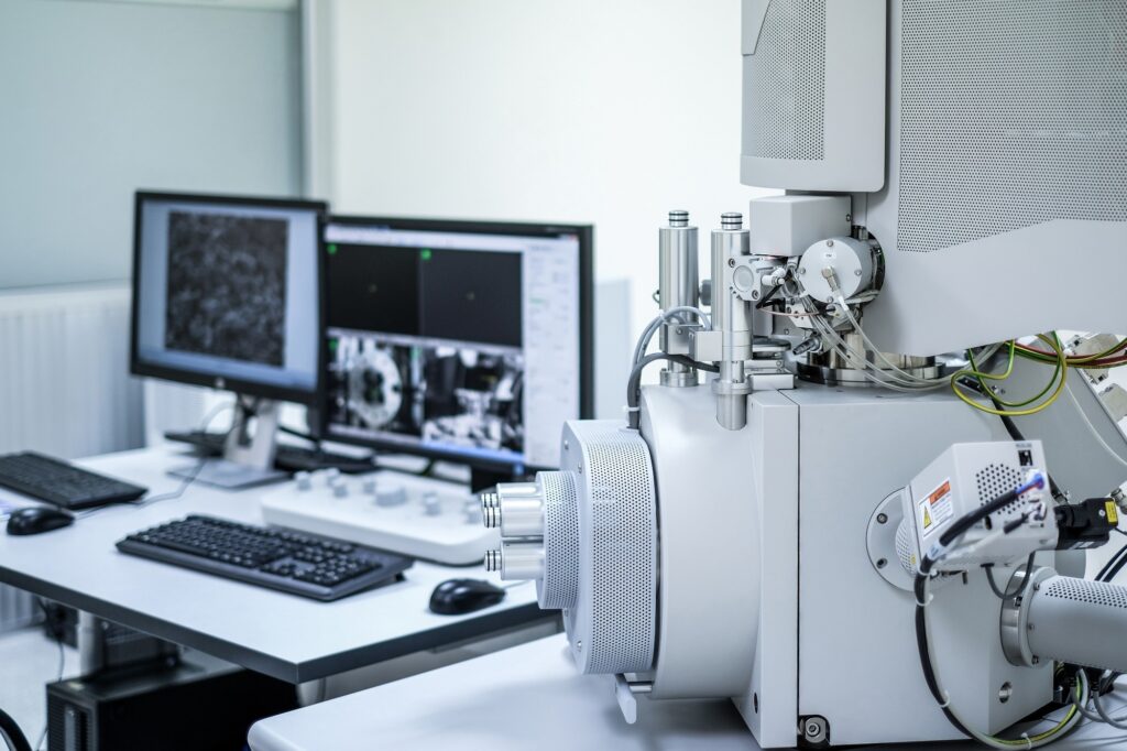
With their high surface area and enhanced physicochemical properties, nanomaterials play a critical role in drug delivery, consumer products, and environmental technologies. However, their nanoscale dimensions enable interactions with cellular components in complex and sometimes unexpected ways, potentially inducing oxidative stress, inflammation, or bioaccumulation. As their use expands, understanding these risks through nanotoxicity testing becomes essential.1
Image Credit: Anucha Cheechang/Shutterstock.com
Why Assess Nanotoxicity?
Assessing nanotoxicity helps ensure the safe use of nanomaterials while protecting human health and the environment. Nanomaterials can enter the body through inhalation, ingestion, or injection. Once inside, they may accumulate in organs and disrupt cellular functions. Their presence in cosmetics, pharmaceuticals, and household goods also raises concerns about environmental exposure. Reliable assessment methods help identify potential hazards before widespread use.2
Methods of Nanotoxicity Assessment
In Vitro Methods
In vitro methods are widely used to assess nanotoxicity through controlled experiments on cell cultures. Cytotoxicity assays such as MTT (tetrazolium-based assays) and LDH (lactate dehydrogenase) release assays evaluate cell viability and membrane integrity.3
Genotoxicity tests, including comet and micronucleus assays, examine DNA damage and chromosomal alterations caused by nanoparticle exposure. By exposing specific cell lines, such as epithelial cells that model the skin, lungs, or gastrointestinal tract, these methods provide critical insights into how nanomaterials interact with different biological barriers.3
For instance, Collins et al. provide key recommendations for conducting in vitro comet assays with mammalian cell cultures. They suggest using non-cytotoxic concentrations, defined as less than 20 % cell viability loss, and recommend concentrations below 100–150 μg/mL for non-cytotoxic nanomaterials.
The selection of cell lines should align with the target organ and exposure route, ensuring relevant biological insights. To capture the full spectrum of nanoparticle interactions, both short-term (2–3 hours) and long-term (24-hour) exposure studies are advised.
Additionally, distinguishing between direct DNA interactions and oxidative stress-induced genotoxicity remains a crucial consideration.4
In Vivo Methods
In vivo studies assess how nanomaterials behave in living organisms. Rodent models help researchers track bioaccumulation and long-term effects on organs like the liver, kidneys, and brain.3 These tests use exposure routes that mimic real-world scenarios, such as inhalation, ingestion, and injection.
While in vivo testing provides valuable data, ethical concerns and species differences highlight the need for alternatives. Regulatory efforts increasingly focus on reducing animal testing by improving in vitro and computational models.3, 5
Computational Methods
Computational toxicology applies in silico models to predict nanotoxicity by analyzing the physicochemical properties of nanoparticles. Techniques such as Quantitative Nanostructure-Toxicity Relationship (QNTR) and Quantitative Structure-Activity Relationship (QSAR) modeling rely on descriptors like particle size, surface charge, aggregation state, and solubility to estimate biological interactions and toxic potential.6
These models offer an efficient alternative to traditional toxicity assessments by reducing dependence on animal studies, minimizing costs, and enabling high-throughput screening. By incorporating data from in vitro experiments, bioinformatics, and machine learning algorithms, computational approaches refine toxicity predictions and enhance our understanding of nanoparticle behavior within biological systems.5
Surface Characterization Techniques
The size, shape, and surface chemistry of nanoparticles influence their interactions with biological systems. Several techniques help researchers analyze these properties:
Scanning Electron Microscopy (SEM): SEM provides high-resolution images of nanoparticles, allowing detailed analysis of their size, shape, and surface morphology. By scanning a focused electron beam across the sample surface, SEM generates images based on the interaction of electrons with the sample. This technique is especially useful for identifying surface features, defects, and coatings.7
Atomic Force Microscopy (AFM): AFM provides three-dimensional imaging and precise measurements of surface properties such as roughness, stiffness, and adhesion strength. Unlike SEM, AFM does not require extensive sample preparation and can operate under ambient or liquid conditions, preserving the native state of nanoparticles. This makes it particularly valuable for studying nanoparticle interactions with biological membranes and their penetration into cells. AFM also quantifies forces between nanoparticles and biological systems, providing insights into their physical interactions and toxicity mechanisms.7
X-Ray Photoelectron Spectroscopy (XPS): XPS is used to analyze the surface chemistry of nanoparticles, including their elemental composition, oxidation states, and surface coatings. This technique is highly sensitive to the outermost layers of nanoparticles, making it ideal for studying functional groups and ligands that influence toxicity.7
Torelli et al. developed an XPS data correction method for non-planar surfaces, improving accuracy when analyzing nanoparticles as small as 20 nm. Such refinements help predict how surface modifications affect biological interactions.8
Protocols for Nanotoxicity Testing
Standardized Guidelines
International organizations like the Organisation for Economic Co-operation and Development (OECD) and the International Organization for Standardization (ISO) have developed guidelines to ensure reliable nanotoxicity testing. The OECD has adapted chemical testing protocols for nanomaterials, addressing issues like aggregation, bioaccumulation, and environmental impact.9
The OECD Sponsorship Programme has assessed various nanomaterials to refine test methodologies, while European initiatives like NANOHARMONY and Gov4Nano focus on standardizing protocols across different regulatory frameworks. These efforts aim to improve test reproducibility and promote global data acceptance under the Mutual Acceptance of Data (MAD) principle.9
Testing Procedures
Nanotoxicity assessments combine in vitro, in vivo, and computational approaches. Testing procedures vary based on exposure routes (oral, dermal, or inhalation) and duration (acute vs. chronic).10
Advanced in vitro assays measure cytotoxicity, oxidative stress, and DNA damage, while in vivo studies track bioaccumulation and organ-specific effects. Newer methods like microfluidic systems and co-culture models enhance test accuracy by mimicking real physiological conditions.10
What Does the Future of Nanotoxicity Testing Look Like?
Despite progress, testing nanotoxicity remains complex. Nanomaterials vary in size, shape, and surface chemistry, making it hard to develop universal protocols. A lack of standardization also leads to inconsistencies across studies.11
Future efforts will focus on integrating advanced technologies. Predictive in silico models and high-throughput in vitro systems will likely play a bigger role in screening nanomaterials. Organ-on-a-chip models could further improve accuracy by replicating human tissue environments.11
To learn more about nanotoxicity testing and advancements in nanomaterial safety, see the resources listed below:
Reference and Further Readings
1. Savage, DT.; Hilt, JZ.; Dziubla, TD. (2019). In Vitro Methods for Assessing Nanoparticle Toxicity. Nanotoxicity: Methods and protocols. https://link.springer.com/protocol/10.1007/978-1-4939-8916-4_1
2. Huang, H.-J.; Lee, Y.-H.; Hsu, Y.-H.; Liao, C.-T.; Lin, Y.-F.; Chiu, H.-W. (2021). Current Strategies in Assessment of Nanotoxicity: Alternatives to in Vivo Animal Testing. International journal of molecular sciences. https://www.mdpi.com/1422-0067/22/8/4216
3. Roberto, MM.; Christofoletti, CA. (2019). How to Assess Nanomaterial Toxicity? An Environmental and Human Health Approach. [Online] IntechOpen. https://www.intechopen.com/chapters/68905
4. Collins, A.; El Yamani, N.; Dusinska, M. (2017). Sensitive Detection of DNA Oxidation Damage Induced by Nanomaterials. Free Radical Biology and Medicine. https://www.sciencedirect.com/science/article/pii/S089158491730062X
5. Budama-Kilinc, Y.; Cakir-Koc, R.; Zorlu, T.; Ozdemir, B.; Karavelioglu, Z.; Egil, AC., Kecel-Gunduz, S. (2018). Assessment of Nano-Toxicity and Safety Profiles of Silver Nanoparticles. [Online] IntechOpen. https://www.intechopen.com/chapters/60486
6. Fourches, D.; Pu, D.; Tassa, C.; Weissleder, R.; Shaw, SY.; Mumper, RJ. Tropsha, A. (2010). Quantitative Nanostructure− Activity Relationship Modeling. ACS nano. https://pubmed.ncbi.nlm.nih.gov/20857979/
7. Gunsolus, IL.; Haynes, CL. (2016). Analytical Aspects of Nanotoxicology. Analytical chemistry. https://pubs.acs.org/doi/full/10.1021/acs.analchem.5b04221
8. Torelli, MD.; Putans, RA.; Tan, Y.; Lohse, SE.; Murphy, CJ.; Hamers, RJ. (2015). Quantitative Determination of Ligand Densities on Nanomaterials by X-Ray Photoelectron Spectroscopy. ACS applied materials & interfaces. https://pubs.acs.org/doi/full/10.1021/am507300x
9. Krug, HF.; Nau, K. (2022). Methods and Protocols in Nanotoxicology. Frontiers Media. https://www.frontiersin.org/journals/toxicology/articles/10.3389/ftox.2022.1093765/full
10. Handy, RD.; van den Brink, N.; Chappell, M.; Mühling, M.; Behra, R.; Dušinská, M.; Simpson, P.; Ahtiainen, J.; Jha, A. N.; Seiter, J. (2012). Practical Considerations for Conducting Ecotoxicity Test Methods with Manufactured Nanomaterials: What Have We Learnt So Far? Ecotoxicology. https://link.springer.com/article/10.1007/s10646-012-0862-y
11. Patel, RJ.; Alexander, A.; Puri, A.; Chatterjee, B. (2021). Current Challenges and Future Needs for Nanotoxicity and Nanosafety Assessment. Nanotechnology in Medicine: Toxicity and Safety. https://onlinelibrary.wiley.com/doi/abs/10.1002/9781119769897.ch14


