Quantum sensing is a rapidly developing field that utilizes the quantum states of particles, such as superposition, entanglement, and spin states, to detect changes in physical, chemical, or biological systems. A promising type of quantum nanosensor is nanodiamonds (NDs) equipped with nitrogen-vacancy (NV) centers. These centers are created by replacing a carbon atom with nitrogen near a lattice vacancy in a diamond structure.
When excited by light, the NV centers emit photons that maintain stable spin information and are sensitive to external influences like magnetic fields, electric fields, and temperature. Changes in these spin states can be detected using optically detected magnetic resonance (ODMR), which measures fluorescence changes under microwave radiation.
In a recent breakthrough, scientists from Okayama University in Japan developed nanodiamond sensors bright enough for bioimaging, with spin properties comparable to those of bulk diamonds. The study, published in ACS Nano, on 16 December 2024, was led by Research Professor Masazumi Fujiwara from Okayama University, in collaboration with Sumitomo Electric Company and the National Institutes for Quantum Science and Technology.
“This is the first demonstration of quantum-grade NDs with exceptionally high-quality spins, a long-awaited breakthrough in the field. These NDs possess properties that have been highly sought after for quantum biosensing and other advanced applications,” says Prof. Fujiwara.
Current ND sensors for bioimaging face two main limitations: high concentrations of spin impurities, which disrupt NV spin states, and surface spin noise, which destabilizes the spin states more rapidly. To overcome these challenges, the researchers focused on producing high-quality diamonds with very few impurities.
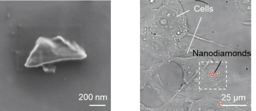
They grew single-crystal diamonds enriched with 99.99% 12C carbon atoms and then introduced a controlled amount of nitrogen (30–60 parts per million) to create an NV center with about 1 part per million. The diamonds were crushed into NDs and suspended in water.
The resulting NDs had a mean size of 277 nanometers and contained 0.6–1.3 parts per million of negatively charged NV centers. They displayed strong fluorescence, achieving a photon count rate of 1500 kHz, making them suitable for bioimaging applications.
These NDs also showed enhanced spin properties compared to commercially available larger NDs. They required 10–20 times less microwave power to achieve a 3% ODMR contrast, had reduced peak splitting, and demonstrated significantly longer spin relaxation times (T1 = 0.68 ms, T2 = 3.2 µs), which were 6 to 11 times longer than those of type-Ib NDs.
These improvements indicate that the NDs possess stable quantum states, which can be accurately detected and measured with low microwave radiation, minimizing the risk of microwave-induced toxicity in cells.
To evaluate their potential for biological sensing, the researchers introduced NDs into HeLa cells and measured the spin properties using ODMR experiments. The NDs were bright enough for clear visibility and produced narrow, reliable spectra despite some impact from Brownian motion (random ND movement within cells).
Furthermore, the NDs were capable of detecting small temperature changes. At temperatures around 300 K and 308 K, the NDs exhibited distinct oscillation frequencies, demonstrating a temperature sensitivity of 0.28 K/√Hz, superior to bare type-Ib NDs.
With these advanced sensing capabilities, the sensor has potential for diverse applications, from biological sensing of cells for early disease detection to monitoring battery health and enhancing thermal management and performance for energy-efficient electronic devices.
“These advancements have the potential to transform health care, technology, and environmental management, improving quality of life and providing sustainable solutions for future challenges,” says Prof. Fujiwara.


