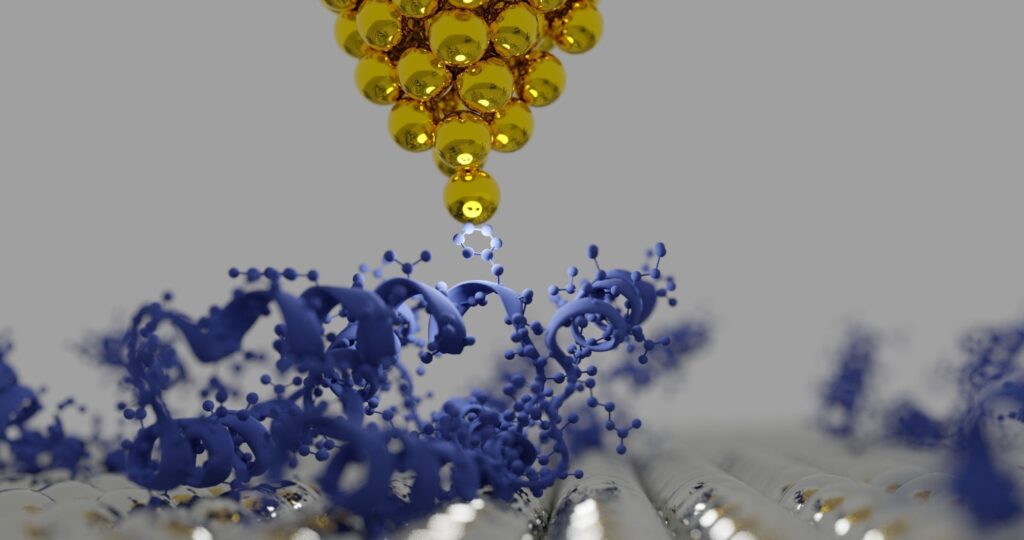
In this interview, Dr. André Körnig, Application Scientist at Bruker BioAFM discusses the applications of BioAFM in the life sciences. He highlights its versatility and ability to image biological samples without extensive preparation.
Could you introduce yourself to our listeners and tell us who you are and what excites you about AFM technologies?
My name is André Körnig, and I’m an application scientist at the BioAFM business unit in Bruker. My interest in AFM technology began during my Master’s thesis, where I realized I was more interested in the life sciences than purely dry material physics. AFM was the first technique I learned, and I got hooked on it. After my PhD, I found an opportunity to work as an application scientist at JPK, and I’ve been enjoying it for 10 years now, exploring different fields and applications. What keeps me excited is the technique’s versatility and constant progression.
What makes AFM technology so special, and why is it important to continue developing it?
I like AFM because it’s quite intuitive; you can imagine it as the finger of a blindfolded person touching surfaces to feel their stiffness and topography. AFM is relatively straightforward to explain and use compared to other complex techniques, especially in biological applications. It allows for examining various biological samples—from small molecules like DNA to entire cells and tissues—in close to native conditions, which is important for biology. The versatility of AFM and its ease of adapting to biological environments make it a valuable tool.
Image Credit: Shutterstock.com/sanjaya viraj bandara
What are some challenges specific to using BioAFM compared to its applications in material sciences?
The biggest challenge is that biological samples are living, which means they change over time. You must ensure the sample stays in place and doesn’t move faster than the scanning can handle. Unlike optical microscopy, where things can freely swim around, AFM requires the sample to be stationary on a surface to be effectively analyzed.
Can you talk about sample preparation, the challenges involved, and some common mistakes you see?
As in all microscopy techniques sample preparation can be crucial. One common mistake is not properly immobilizing the sample, which can cause it to move during scanning. For instance, I faced challenges measuring magnetotactic bacteria during my PhD because they wouldn’t stay still long enough for analysis. In such cases the sample preparation requires an understanding of chemistry for surface modifications, the biological behavior of the sample, and an interdisciplinary approach to properly balance all these aspects. But in general sample preparation for AFM is quite easy, e.g. compared to Electron Microscopy.
Conversations on AFM #2: Applications of BioAFM in Life Science
Video Credit: Bruker BioAFM
What new applications or trends have you seen in AFM over the past decade?
There has been significant development in terms of speed. Imaging used to take 10 minutes per image, but now it can be much faster, such as multiple images per second. This opens new possibilities for studying dynamics. Additionally, mechanical analysis—determining stiffness and elasticity—has evolved significantly, especially in fields like cancer research. Another major trend is making the system easier to use, so more people, even those without deep technical knowledge, can perform sophisticated measurements.
Can you tell us about the multi-parametric imaging of fibroblasts and what it involves?
Fibroblasts cells are quite robust and easy to work with, and a great model system for general imaging. Multi-parametric imaging means seeing the topography of the cells and measuring other properties like stiffness, adhesion, or even internal structures. We poke into the sample to measure how it reacts, which allows us to obtain mechanical properties like Young’s modulus. Additionally, we can attach molecules to the cantilever to measure interactions directly while imaging.
Could you explain the TappingMode and PeakForce TappingMode specifically for Bruker Nano?
TappingMode involves oscillating the cantilever at its resonance frequency, which is great for fast imaging. PeakForce TappingMode, a special development by Bruker, allows access to quantitative force curves while imaging, giving us data like indentation forces and stiffness. It’s particularly useful in polymer sciences and other materials research.
What about the Quantitative Imaging (QI) mode? How is it different, and when is it most useful?
The QI mode was originally developed by JPK and is now part of Bruker. It’s easy to use and less likely to disturb or move the sample, making it ideal for delicate or poorly fixed samples. It’s intuitive to set up and provides quantitative data, making it my go-to mode if I’m unsure about a sample’s characteristics. It’s slower than other modes, but it’s very stable and ideal for exploration and precise analysis.
What do you think is the future direction for AFM in the next five years?
I think AFM will continue to become more robust and easier to use, making it more accessible outside research institutions and possibly into medical centers. There is great potential in cancer research and other medical applications, where AFM’s ability to measure mechanical properties in native conditions could prove incredibly useful. The development focus will be on making it more reliable for users who aren’t necessarily experts in the technology.
Click Here to Listen to the Podcast
About Dr. André Körnig
Dr. André Körnig is a biophysicist who did his PhD at the Max Planck Institute of Colloids and Interfaces in Potsdam Golm, Germany. He is a Senior Application Scientist at Bruker BioAFM and responsible for carrying out customer training and installations worldwide. His work focuses on the biophysical applications of AFM, such as imaging of single cells and biomolecules, nanomechanical analysis using force spectroscopy, indentation, and microrheology, and the investigation of dynamic mechanisms using high-speed AFM imaging.
This information has been sourced, reviewed and, adapted from materials provided by Bruker BioAFM.
For more information on this source, please visit Bruker BioAFM.
Disclaimer: The views expressed here are those of the interviewee and do not necessarily represent the views of AZoM.com Limited (T/A) AZoNetwork, the owner and operator of this website. This disclaimer forms part of the Terms and Conditions of use of this website.


