


Fluorescent nanobeads offer great potential for many applications in both basic and applied research.
Recently, scientists have been offered a wide range of technical solutions in fluorescence imaging, enabling significant advancement in fields such as microscopy and diagnostics. However, these optical markers mainly covered the microscale spectrum and/or faced limitations in optical performance and adaptability.
The recent progress made in the field of nanoscience has allowed for the creation of precisely controlled nanostructures with customized optical properties, unveiling entirely unexplored opportunities and intriguing possibilities across various research domains.
The newly devised fluorescent systems amalgamate diminutive dimensions (typically <100 nm) with elevated brightness, photostability, adjustable spectral features/surface charge/chemistry, and biocompatibility.
These distinctive properties open up novel applications in areas where size plays a crucial role, including cellular studies, high-resolution microscopies, drug delivery, and sensing.
Possible applications of nanobeads include:
- Fluorescence/Confocal microscopy (including super-resolution techniques)
- Flow cytometry
- Imaging, sensing, and diagnostics
- Nanomedicine and nanotoxicology
Fluorescent Nanobeads
The bright and stable fluorescence emitters with nanoscale dimensions open many stimulating opportunities in optical imaging.
For example, high-resolution applications that demand particle imaging, even at the single-particle level, become a standard practice rather than a formidable task and can be routinely executed by non-specialized researchers and/or commercially available instruments and devices.
This is a key point, as the availability of a fluorescent tag that is highly bright and functionalizable with many (bio)molecules enables the development of many applications in imaging, sensing, and diagnostics.
Cellular/tissue imaging can also significantly benefit from the use of fluorescent nanobeads, as their brightness and non-cytotoxicity enable high-resolution intracellular localization and tracking of targets of interest in both fixed and living cells.1
The nanobeads’ extreme photostability in biological environments allows for prolonged studies, exhibiting minimal or no photobleaching even after extended irradiation.
The advantageous characteristics of photostability and brightness can be harnessed in super-resolution imaging techniques, such as in Stimulated Emission Depletion (STED) microscopy.
The STED technique, pioneered by 2014 Nobel Laureate Stefan W. Hell, surpasses the diffraction limit of optical microscopes (approximately 200 nm) by selectively deactivating fluorophores, facilitating a substantial enhancement of lateral resolution (down to 30-40 nm with commercial microscopes) in fluorescence images.
This important advancement in imaging has recently led to many outstanding findings in biology and biophysics.2,3 Merck offers well-characterized fluorescent nanobeads (Figure 1), which allow super-resolution STED imaging, permitting a strong push in lateral resolution down to 30 nm.
In these applications, the accurate size control and monodispersion provided by this material enabled reliable imaging of the real dimensions of the nanobeads. Super-resolved images can also be obtained in the biological environment, including cellular imaging (Figures 2 & 3).
Figure 1. Left: Multicolor fluorescent nanobeads dispersed in water (emission is available from blue to red/infrared). Right: Representative TEM image of monodispersed silica nanobeads (Prod No. 797952). Image Credit: Merck
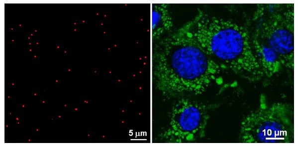
Figure 2. Confocal microscopy image of 120 nm fluorescent nanobeads (Prod No. 797863) on a glass slide (Left), or internalized by A549 cells (Right). A very bright and stable signal is generated by the nanobeads. The biocompatibility and customizable surface chemistry allows successful applications, such as biological imaging and targeted drug delivery. Image Credit: Merck
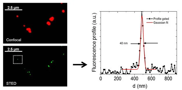
Figure 3. Confocal and super-resolution gSTED microscopy image of 25 nm fluorescent nanobeads (Prod No. 797901). The high brightness and photostability of the nanobeads easily allow a strong improvement in lateral resolution. In these applications, an accurate control of the particle size is crucial, and our nanobead products offer the best quality, enabling superior imaging. The biocompatibility of the nanobeads opens up a wealth of applications in both in-vitro and in-vivo systems. Image Credit: Merck
Another vital application of fluorescent nanobeads is evident in flow cytometry. The nanobeads’ high brightness facilitates the easy detection of biological processes and cellular interactions using standard instruments.
Passive cellular internalization and specific targeting can be monitored using flow cytometry, including in multicolor assays. The nanobeads can be functionalized with antibodies or aptamers, facilitating targeted applications and diagnostics.
These nanobeads have been instrumental in the sensitive and rapid detection of cancer cells via flow cytometry, surpassing the sensitivity of standard methods.4
Additionally, a highly sensitive and specific method for detecting Staphylococcus aureus was achieved by He et al.5 Nanomedicine and nanotoxicology investigations greatly benefit from the use of these bright nanobeads (Figure 4).
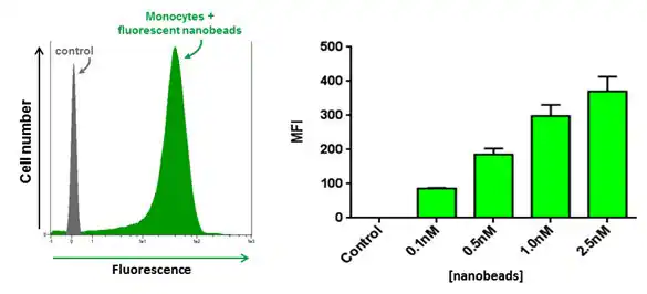
Figure 4. Internalization of our fluorescent nanobeads in human primary CD14+ monocytes as probed by flow cytometry. Meaning fluorescence intensity increases as a function of nanobeads concentration. Image Credit: Merck
Summary
In these applications, the effectiveness and adaptability of the fluorescent probes are crucial parameters.
Merck’s Material Science portfolio encompasses various fluorescent nanobeads ranging in size from 25 to 250 nm.
These ensure precise control over particle uniformity, fluorescence properties (from blue to infrared emission), and surface charge and chemistry (e.g., carboxylic, amino, and sulfonic acid groups) to facilitate appropriate biological interactions or specific bioconjugation for targeting.
All of Merck’s fluorescent nanobeads are highly bright and photostable and are characterized by high biocompatibility and sterility.
References
- Chu Z, Huang Y, Tao Q, Li Q. 2011. Cellular uptake, evolution, and excretion of silica nanoparticles in human cells. Nanoscale. 3(8):3291. https://doi.org/10.1039/c1nr10499c
- Westphal V, Rizzoli SO, Lauterbach MA, Kamin D, Jahn R, Hell SW. 2008. Video-Rate Far-Field Optical Nanoscopy Dissects Synaptic Vesicle Movement. Science. 320(5873):246-249. https://doi.org/10.1126/science.1154228
- Eggeling C, Ringemann C, Medda R, Schwarzmann G, Sandhoff K, Polyakova S, Belov VN, Hein B, von Middendorff C, Schönle A, et al. 2009. Direct observation of the nanoscale dynamics of membrane lipids in a living cell. Nature. 457(7233):1159-1162. https://doi.org/10.1038/nature07596
- Estévez M-, O?Donoghue MB, Chen X, Tan W. 2009. Highly fluorescent dye-doped silica nanoparticles increase flow cytometry sensitivity for cancer cell monitoring. Nano Res.. 2(6):448-461. https://doi.org/10.1007/s12274-009-9041-8
- He X, Li Y, He D, Wang K, Shangguan J, Shi H. 2014. Aptamer-Fluorescent Silica Nanoparticles Bioconjugates Based Dual-Color Flow Cytometry for Specific Detection of Staphylococcus aureus. Journal of Biomedical Nanotechnology. 10(7):1359-1368. https://doi.org/10.1166/jbn.2014.1828

This information has been sourced, reviewed and adapted from materials provided by Merck.
For more information on this source, please visit Merck.
