Wounds that are superficial for some can be life-threatening for others. With diabetic wounds, healing can be slow, particularly in the feet, increasing the tissue’s susceptibility to infection. Foot ulcers and other diabetic foot complications have similar mortality rates to some cancers, yet progress toward improved treatments has plateaued. Now, researchers may have found a better way to kickstart the healing process.
Arizona State University (ASU) bioengineers have developed a multistep strategy that applies different nanomaterials to wounds at different times to support both early- and late-stage healing. In a study published in the journal Biomaterials, the authors’ method outperformed a common wound dressing in a diabetic mouse model, closing wounds faster and producing more robust skin tissue.
“Healing a wound is like building a house. You have to lay the foundation first before you can put in the plumbing,” said co-first author Jordan Yaron, Ph.D., a bioengineering assistant research professor at ASU. “With our approach, we’re mindful of which stage the wound is in. Providing the right treatment at the right time is key.”
The researchers’ analysis also suggests that their approach unexpectedly activated an immune cell population not normally seen in wounds that can resolve inflammation, which highlights a new potential avenue to accelerate healing.
A multifaceted solution for a multifaceted problem
Clinically, the standard practice for wounds is to keep them clean and use a dressing to protect them while they heal. This approach gets the job done for most injuries but falls short for patients with conditions that interfere with the healing process, such as diabetes. In addition to causing poor circulation and neuropathy, diabetes can disrupt wound healing by impairing the function of various immune cells.
The researchers devised a strategy to treat wounds like these and compared it to a commonly used dressing in a diabetic mouse model.
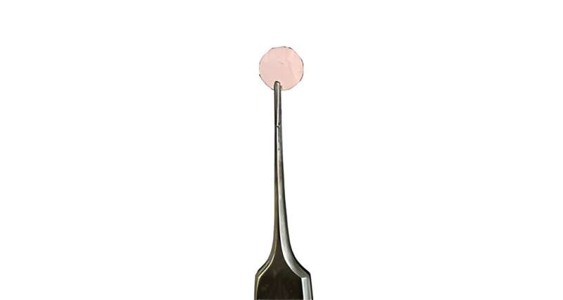
For the first step, the team fabricated a silk nanomaterial dressing embedded with gold nanorods. Because gold nanoparticles readily convert light to heat, the team was able to direct a laser at dressings placed over fresh wounds in mice, producing heat that quickly sealed them in place and provided a high level of protection.
The strategy, which the authors previously found success with, creates something akin to an instantaneous scab, Yaron explained. This time around, the authors added histamine to the mix, a natural biochemical produced by the immune system that plays important roles in inflammation, blood vessel development, and allergic reactions.
Inflammation dominates the body’s initial response to injuries, but eventually subsides to allow the body to rebuild. However, diabetic wounds can get stuck in first gear, maintaining persistent, low-grade inflammation, which can inhibit the healing process.

“Since the wound is stalled, we wanted to co-deliver histamine with the dressing, to give a push and bring the inflammation stage to a resolution. Then we could introduce another strategy to take care of the subsequent phases of healing,” said senior and corresponding author Kaushal Rege, Ph.D., a chemical engineering professor at ASU.
The authors monitored the wounded mice for 11 days and found that animals treated with a combination of the nanomaterial dressing and histamine healed at the fastest rate compared to those treated with the standard dressing with histamine or the nanomaterial dressing alone.
The researchers mechanically tested the healed skin as well, finding that the tissue treated with both the nanomaterial and histamine was strongest and most similar to unwounded skin.
How exactly did the treatment speed up the process so drastically? For a better understanding, the team analyzed tissue samples by assessing gene expression and examining cells under the microscope.
They found that a specific immune cell type, N2 neutrophils, were highly prevalent in treated wounds up to seven days post-injury. As the first responders of the immune system, these cells usually clear out within a day or two, making their presence in the wound after a week highly unusual. But since they are known to produce histamine and other reparative molecules, it is also possible that these immune cells are the linchpin of the treatment, noted Yaron.
The team plans to dig deeper into the cause of the neutrophils’ presence as well as their role in wound healing.
The investigators’ next step in the study was to see if they could improve healing further by accelerating the post-inflammation phase wherein cells proliferate and remodel skin tissue.
In a second set of mice, the authors followed up their initial treatment with a pair of nanoparticles they previously developed that were derived from two particular growth factors—proteins native to the body that promote the formation of skin tissue.
The team injected the nanoparticles at various time points into the nanomaterial-dressed wound bed, finding that delivery on day six had the best outcomes with regards to wound closure and tissue strength. This time point corresponds to a transitionary phase in which cells begin proliferating and remodeling tissue.
With promising results in mice behind them, the authors are now testing their strategy in larger animal models more relevant to human health, such as pigs.
“The authors put together previously existing components in a unique way. By considering the timing of bioactive delivery and analyzing the immune response, the team put forth an impressive study. I believe they are in a good position moving forward,” said David Rampulla, Ph.D., director of the Division of Discovery Science and Technology at the National Institute of Biomedical Imaging and Bioengineering (NIBIB).


