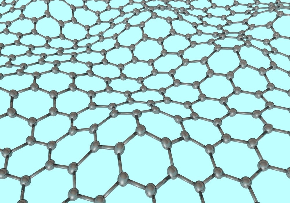
Graphene oxide (GO) nanoflakes are two-dimensional (2D) nanomaterials with unique properties, such as high biocompatibility and exceptional conductivity, and are used in several applications. Here, we discuss the different methods used for the characterization of GO nanoflakes.
Image Credit: Jarek_Sz/Shutterstock.com
GO Nanoflake Characterization Methods
GO primarily refers to an oxidized form of graphene that is hydrophilic and dispersible in water. GO nanoflakes possess high surface area, excellent mechanical strength, and superior optical properties, which make them suitable for energy storage, biomedical devices, sensors, drug delivery, membranes and composites, optoelectronics, and solar cells.
Atomic force microscopy (AFM), scanning electron microscopy (SEM), transmission electron microscopy (TEM), Raman spectroscopy, and X-ray diffraction (XRD) are commonly used for the characterization of GO nanoflakes, including their thickness, surface roughness, internal structure, surface morphology, and crystal structure.
AFM
AFM is a non-optical high-resolution imaging technique that allows non-destructive and accurate measurements of a sample surface’s mechanical, optical, chemical, magnetic, electrical, and topographical properties with a very high resolution in ultrahigh vacuum, liquids, or air.
AFM is used extensively for GO flake characterization as AFM instrument is less expensive to operate and purchase than instruments used in other characterization methods, such as TEM.
In a paper published in the journal Toxicology in Vitro, researchers used AFM to characterize the size and morphology of the single and four-layer GO nanoflakes. They obtained AFM images from the flakes deposited on silicon dioxide/silicon wafers. The thickness of the single and four-layer GO nanoflakes determined using AFM was approximately one nm and three nm, respectively.
Additionally, the length distribution of the GO flakes was between one and 25 μm, with the mean length for the one- and four-layer GO ranging from one to 15 μm. The GO length distribution was determined after analyzing 150 nanostructures and considering the longest edge of the flake as its length.
In a paper published in the journal Nanotechnology, an international interlaboratory comparison was performed on GO flake thickness measurements using AFM. Twelve laboratories participated in this project to improve the thickness measurement equivalence for 2D flakes.
The participants used AFM to image a total of 10 flakes and then measured the thickness of every flake. A detailed protocol, including analysis and imaging methods, was used by all participants.
Participants calibrated the AFM scanner Z-axis by imaging a proper step-height standard, such as a certified reference material (CRM), and employed intermittent contact mode for imaging using intermittent-contact mode cantilevers with 40 N m−1 force constant, 8-12 nm probe apex size, and 300 kHz resonant frequency.
The scan parameters were optimized and every participant scanned a 20 μm × 20 μm area, followed by higher resolution imaging of ten individual flakes at a higher pixel density of 512 pixels × 512 pixels and using smaller scan sizes of 2 μm × 2 μm/5 μm × 5 μm. The results of the twelve participants demonstrated a 0.93 nm average flake thickness with a 0.08 nm standard deviation.
TEM
TEM is an analytical technique utilized to visualize the smallest structures in matter. The technique can reveal minute details, including the arrangement of atoms, the presence of defects, and the elemental composition, at the atomic scale by magnifying the nanometer structures up to 50 million times, as electrons can possess a significantly shorter wavelength compared to visible light when they are accelerated through a strong electromagnetic field.
In a paper published in the journal Electrochimica Acta, researchers synthesized reduced GO nanoflakes (RGONF) from graphite using a simple one-step wet chemical approach by a thermal reduction at low temperature.
Several methods were used to characterize the synthesized RGONF, including XRD, Raman spectroscopy, X-ray photoelectron spectroscopy, TEM, and field emission scanning electron microscopy (FE-SEM). The SEM and TEM images demonstrated the successful synthesis of RGONFs.
In another paper published in the journal, researchers developed a novel one-step method for synthesizing GO nanostructures using pulsed laser ablation in GO solution. SEM and TEM were used to demonstrate the formation of different GO nanostructure shapes, such as ribbons, quantum dots, and nanoflakes, including nano-disks, nano-hexagons, nano-triangles, nano-rectangles, and nano-squares. Results from the TEM and SEM studies indicated successful synthesis of various GO nanostructures using the proposed method.
SEM
SEM is a highly versatile technique utilized to obtain detailed surface information and high-resolution images of samples. The technique employs a focused beam of electrons to scan the specimen surface and generate images at significantly higher resolution than optical microscopy. The SEM instrument resolution can range from less than one nm to several nanometers.
SEM has been used to characterize different GO nanostructures, including GO nanoflakes, in a study published in the journal Nanoscale. SEM images indicated the shapes of GO nanoflakes with a few tenths or hundreds of nanometers in size after 10 and 15 min irradiation time.
The GO nanoflakes produced using the pulsed laser ablation method include nano-disks, nano-triangles, nano-rectangles, and nano-squares. However, smaller GO nanostructures synthesized after over 20 min of irradiation time could not be effectively characterized using SEM.
Raman Spectroscopy
Raman spectroscopy is a non-destructive chemical analysis technique used to obtain detailed information about molecular interactions, crystallinity, phase and polymorphy, and chemical structure. The light scattering technique depends on the interaction between light and chemical bonds in a material.
In Raman spectroscopy, a molecule scatters the incident light from a high-intensity laser light source.
Most of the light is scattered at the same wavelength as the laser source and does not provide any crucial information about the material. However, an extremely small amount of light scattered at different wavelengths depending on the chemical structure of the material, which is known as Raman Scatter, provides critical information about GO nanoflakes.
In a study published in the journal Electrochimica Acta, researchers employed Raman spectroscopy at 758 nm excitation wavelength to characterize RGONF synthesized using a simple one-step wet chemical approach.
The Raman spectrum for RGONF displayed a G band peak at 1572 cm-1, indicating a graphitized structure formation, and a D band peak at 1308 cm-1 corresponding to the disorder-induced phonon mode.
Additionally, the G band peak intensity was significantly higher than the D band peak intensity. The G band peak was accompanied by a shoulder peak at 1602 cm-1 due to graphene edges and finite-size graphite crystals.
Moreover, the 2D band peak at 2616 cm-1 confirmed the presence of graphene, while the D’ band peak indicated the presence of graphene edges and defects and a more nanocrystalline structure, which are the common features of RGONF.
See More: Chemical Reduction of Graphene Oxide (rGO)
References and Further Reading
Bu, T., Gao, H., Yao, Y., Wang, J., Pollard, A., Legge, E., Clifford, C., Delvallee, A., Ducourtieux, S., Lawn, M., Babic, B., Coleman, V., Jamting, A., Zou, S., Chen, Maohui., Jakubek, Z., Iacob, E., Chanthawong, N., Mongkolsuttirat, K., Ren, Lingling. (2023). Thickness measurements of graphene oxide flakes using atomic force microscopy: Results of an international interlaboratory comparison. Nanotechnology. https://doi.org/10.1088/1361-6528/acbf58.
Peruzynska, M., Cendrowski, K., Barylak, M., Tkacz, M., Piotrowska, K., Kurzawski, M., Mijowska, E., Drozdzik, M. (2017). Comparative in vitro study of single and four layer graphene oxide nanoflakes — Cytotoxicity and cellular uptake. Toxicology in Vitro, 41, 205-213. https://doi.org/10.1016/j.tiv.2017.03.005
Rezaei, B., Jahromi, A. R. T., Ensafi, A. A. (2016). Ni-Co-Se nanoparticles modified reduced graphene oxide nanoflakes, an advance electrocatalyst for highly efficient hydrogen evolution reaction. Electrochimica Acta, 213, 423-431. https://doi.org/10.1016/j.electacta.2016.07.133
Lin, T. N., Chih, K. H., Yuan, C. T., Shen, J. L., Lin, C. A. J., Liu, W. R. (2015). Laser-ablation production of graphene oxide nanostructures: From ribbons to quantum dots. Nanoscale. https://doi.org/10.1039/c4nr05737f.
What is Atomic Force Microscopy (AFM) [Online] Available at https://www.nanoandmore.com/eu/what-is-atomic-force-microscopy
Transmission Electron Microscopy [Online] Available at https://www.nanoscience.com/techniques/transmission-electron-microscopy/
Scanning Electron Microscopy [Online] Available at https://www.nanoscience.com/techniques/scanning-electron-microscopy/
What is Raman Spectroscopy? [Online] Available at https://www.horiba.com/usa/scientific/technologies/raman-imaging-and-spectroscopy/raman-spectroscopy/
Graphene Oxide Nanoflakes May Be A Potential Ally Against Alzheimer’s [Online]


