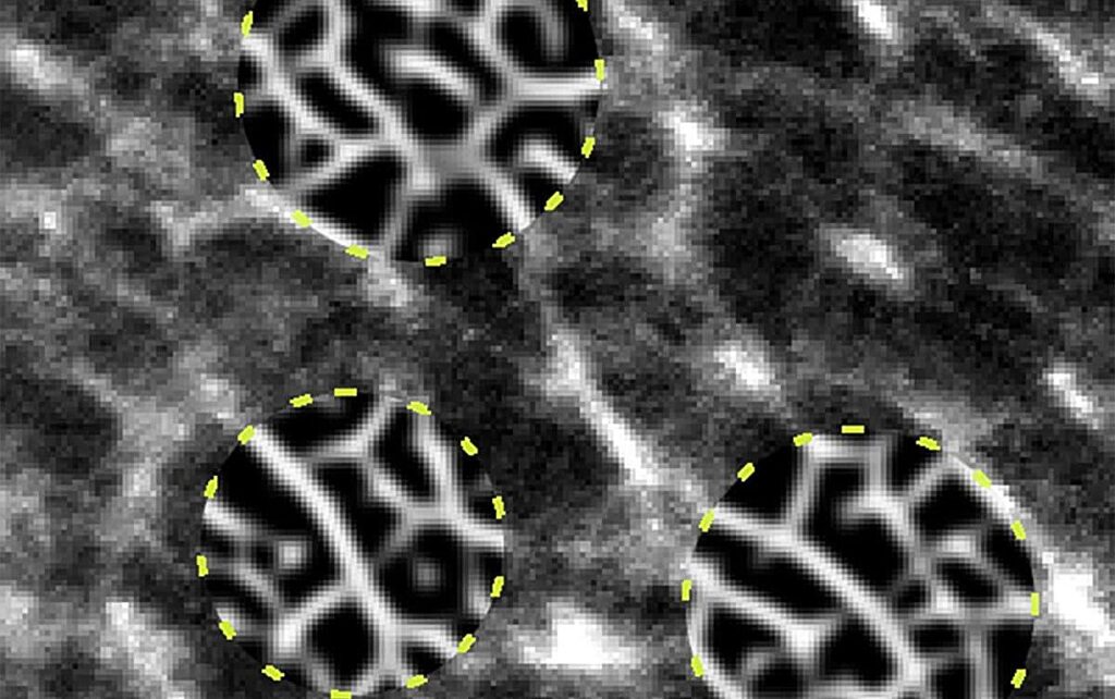
Researchers at University of Tsukuba have developed a new imaging method that clearly visualizes nanoscale structures within rubber materials. The study is published in the journal ACS Applied Nano Materials.
Conventional electron microscopy often produces noisy images that obscure rubber’s internal contours. The proposed method successfully captures the mesh-like molecular network structure and quantifies factors involved in the internal structure.
Rubber possesses unique properties such as softness and stretchability, which are exploited in applications ranging from tires to medical materials. Molecular bonding forms a complex network structure that considerably influences the physical properties of rubber. However, the precise internal structure of rubber is not easily discerned in conventional electron microscope images because the outlines are obscured by noise.
To address this issue, researchers have developed a new image-processing method for electron microscope images that selectively enhances the visibility of areas in which rubber molecules aggregate into network-like structures.
By integrating knowledge of the rubber material with advanced mathematical techniques, the new method visually clarifies the internal network structure of rubber at the nanoscale, even in very noisy electron microscope images with unclear outlines.
The network region, which must be identified manually by conventional methods, can be calculated automatically by the new method, thereby eliminating the need for arbitrariness and enabling simultaneous analysis of multiple samples.
The researchers measured the network length of each sample using the new method. The processed data were highly correlated with the experimental values, thus confirming the new method’s reliability.
The findings of this research are expected to drive the development of safe, economical, and high-performance rubber materials, contributing to societal benefits such as resource and energy conservation.


